
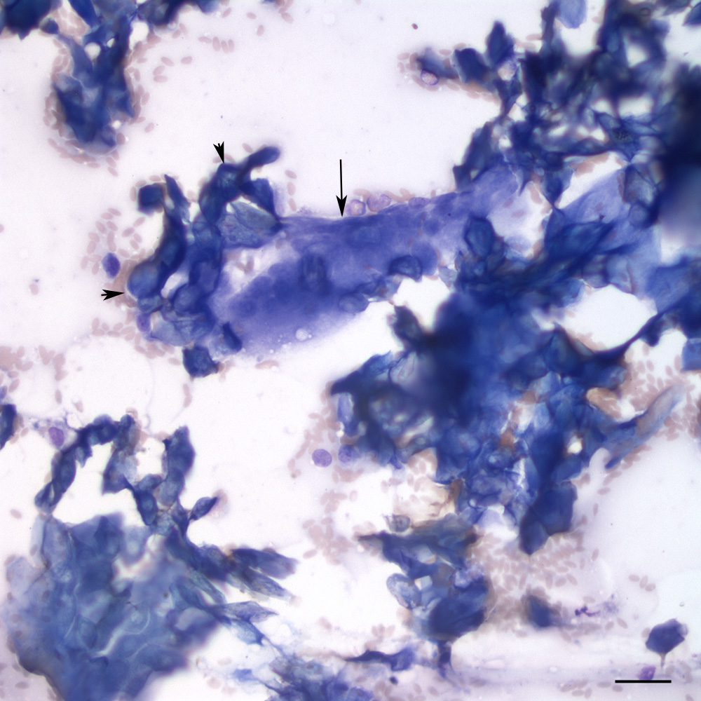
The smears contained sheets of dark blue anucleated keratinized squamous cells, many of which had central clearing in the location of the nucleus (arrowheads), so-called ‘ghost’ or ‘shadow’ cells. These cells usually arise from the matrical bulb of the hair follicle, indicating the mass originates from a hair follicle. There were also multinucleated giant cells (arrow) (modified Wright’s stain, scale bar = 25 um)