
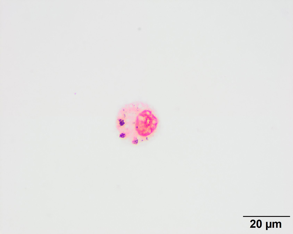
This image shows a macrophage containing 3 small aggregates of blue coarse pigment (positive staining reaction), indicating the presence of iron as hemosiderin in the cytoplasm. This stain confirms that the brown pigment in the macrophages in the modified Wright’s stain was hemosiderin (Prussian blue, 100x objective).