
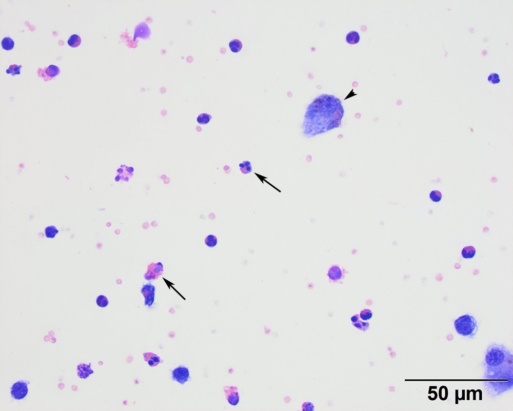
The smear contains many eosinophils (black arrows) with a few macrophages. One macrophage contains small brown globules (hemosiderin, presumptive) and phagocytized eosinophil granules (arrowhead). There is a small amount of blood in the background (Wright’s, 50x objective).