
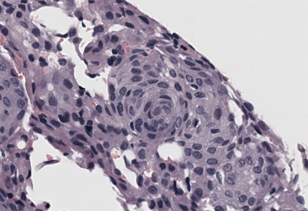
A higher power view of the mass illustrates the whorls and the central to eccentric round to oval to elongate nuclei with stippled to vesicular chromatin and single nucleoli. The cells have a moderate amount of amphophilic cytoplasm with indistinct borders. Their spindled shape is more obvious in the whorl (hematoxylin & eosin, high magnification)