
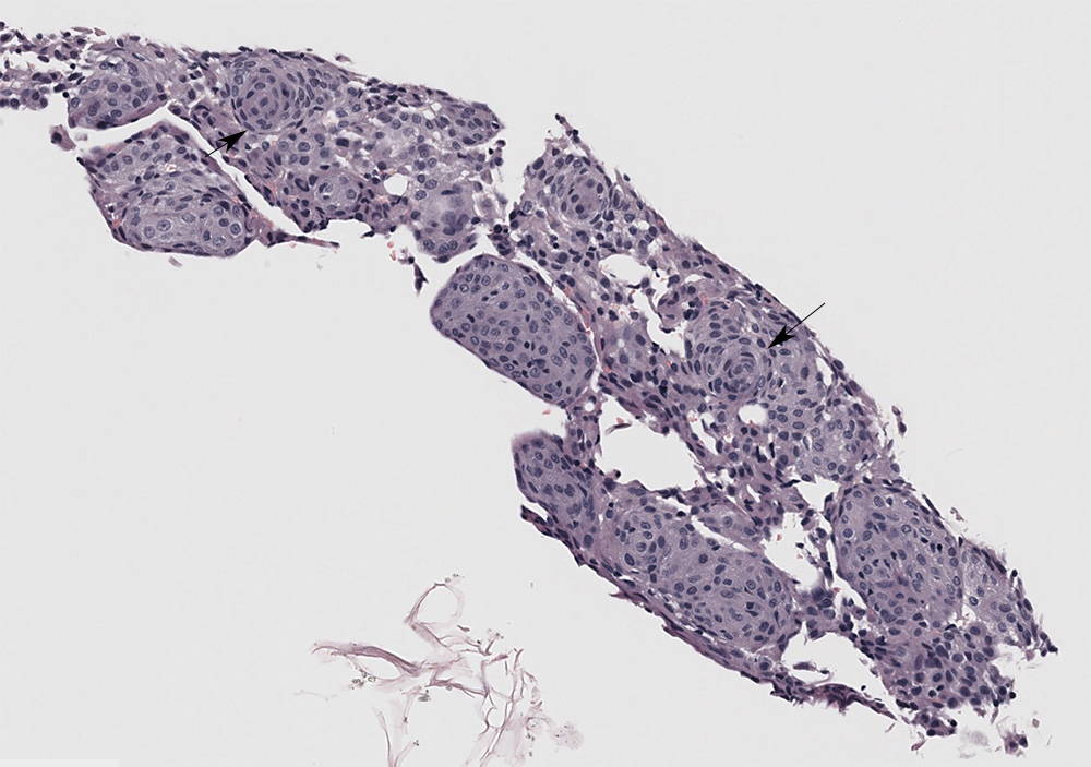
The Tru-cut biopsy had minor crush artifact on the edges. The mass consisted of spindled to polygonal cells with indistinct boundaries arranged in circular to whorling (arrows) arrangements with a few flat sheets (hematoxylin & eosin stain, low magnification)