
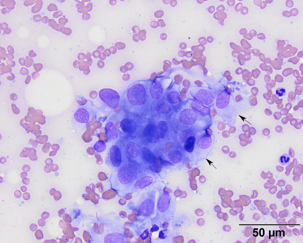
The aspirate consisted of aggregates of spindled to polygonal cells, with round to oval nuclei containing coarsely stippled chromatin. Note, the tapering boundaries (arrows). Nucleoli are indistinct. In this aggregate, the cells display mild to moderate anisokaryosis (modified Wright’s stain)