
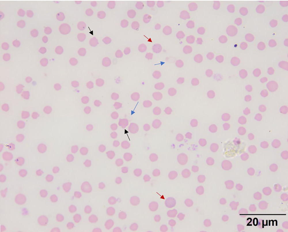
Note the marked anisocytosis, which is due to macrocytes and polychromatophils, and basophilic stippling in polychromatophils (red arrows). Heinz bodies are difficult to see on the intact red blood cells, forming small light red to colorless protrusions on the surface of the cells (black arrows). The Heinz bodies are more distinct (dark red circular structures) on the ghost red blood cells (blue arrows) (Wright’s stain, 100x objective). There are also single Howell-jolly bodies (nuclear remnants) in several red blood cells (e.g. in the two cells to the left of the upper black arrow). Goat red blood cells are quite small normally so these changes can be difficult to see, even with the 100x objective.