
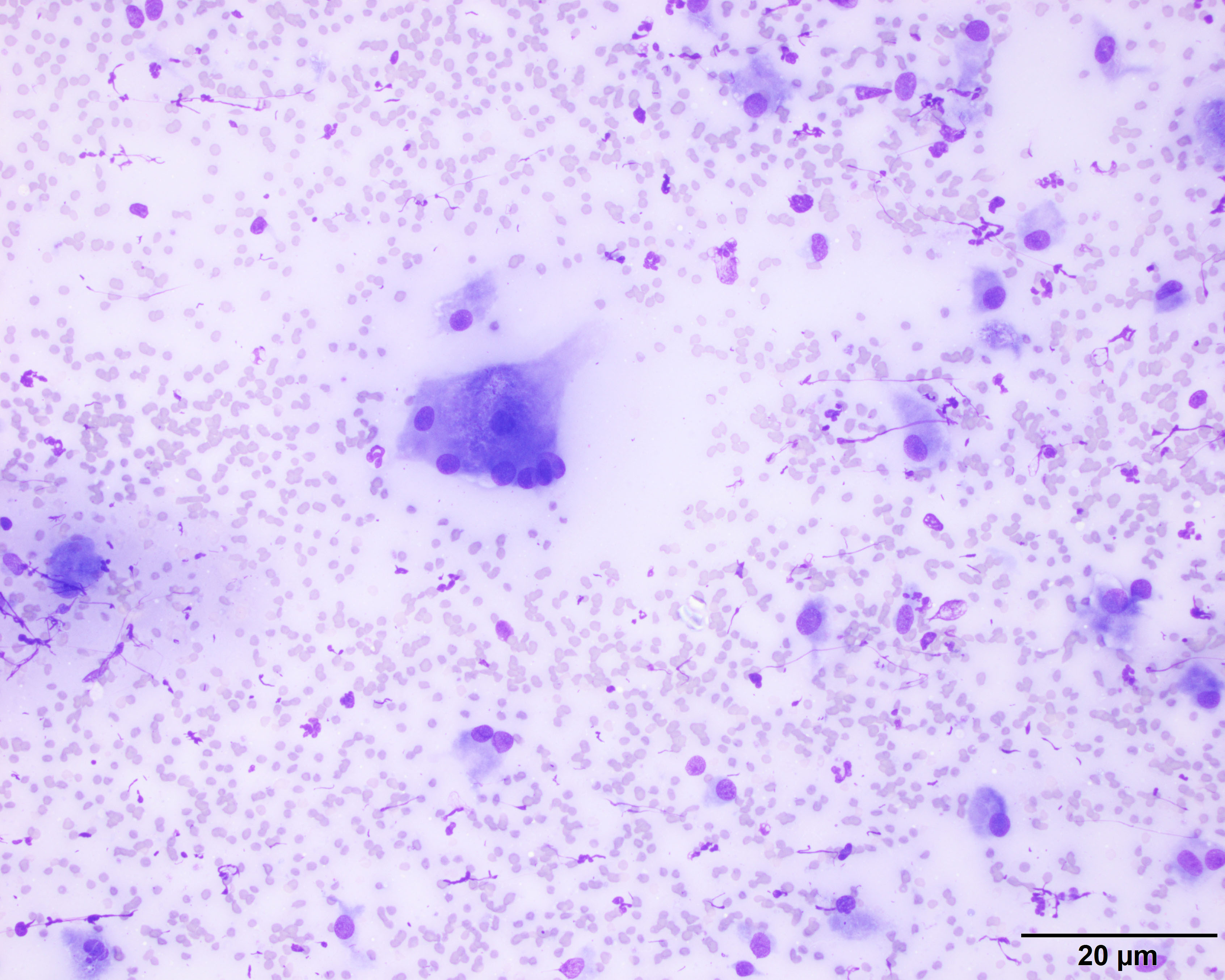
The aspirate of the mass revealed moderate numbers of epithelioid macrophages and degenerate neutrophils within a background of moderate amounts of blood. There is a multinucleated giant cell with phagocytized organisms in the center (20x objective, Wright’s stain).