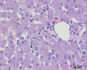
There are moderate numbers of red-brown intracytoplasmic granules within hepatocytes (arrow) in the centrilobular region (zone 3). Macrophages containing copper or cellular debris form small aggregates in this region (arrowhead). Low numbers of small lymphocytes are seen around the central vein (middle right), indicating a mild centrilobular hepatitis (lymphoplasmacytic, overall) (H&E stain).

