
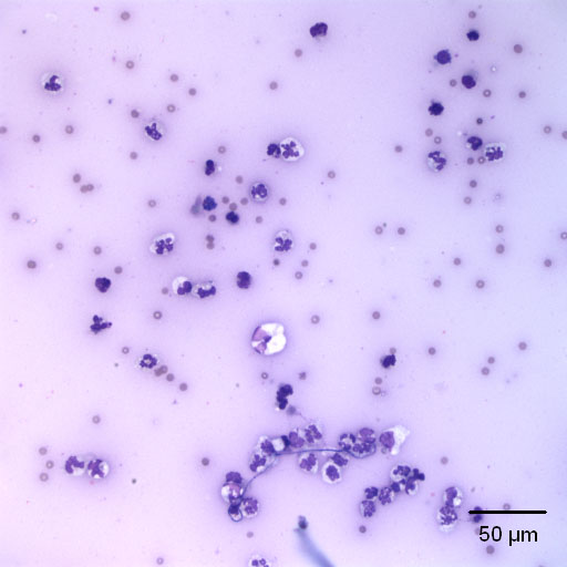
The fluid has increased cellularity, consisting primarily of neutrophils some of which are markedly degenerat (as seen by the swollen cell in the center). The cells are also not windrowing (lining up), supporting decreased viscosity (Wright’s stain). These are abnormal findings, since in a normal joint fluid neutrophils should not exceed the 10% of the cell population and the nucleated cell count is usually <1,000 cells/uL.