
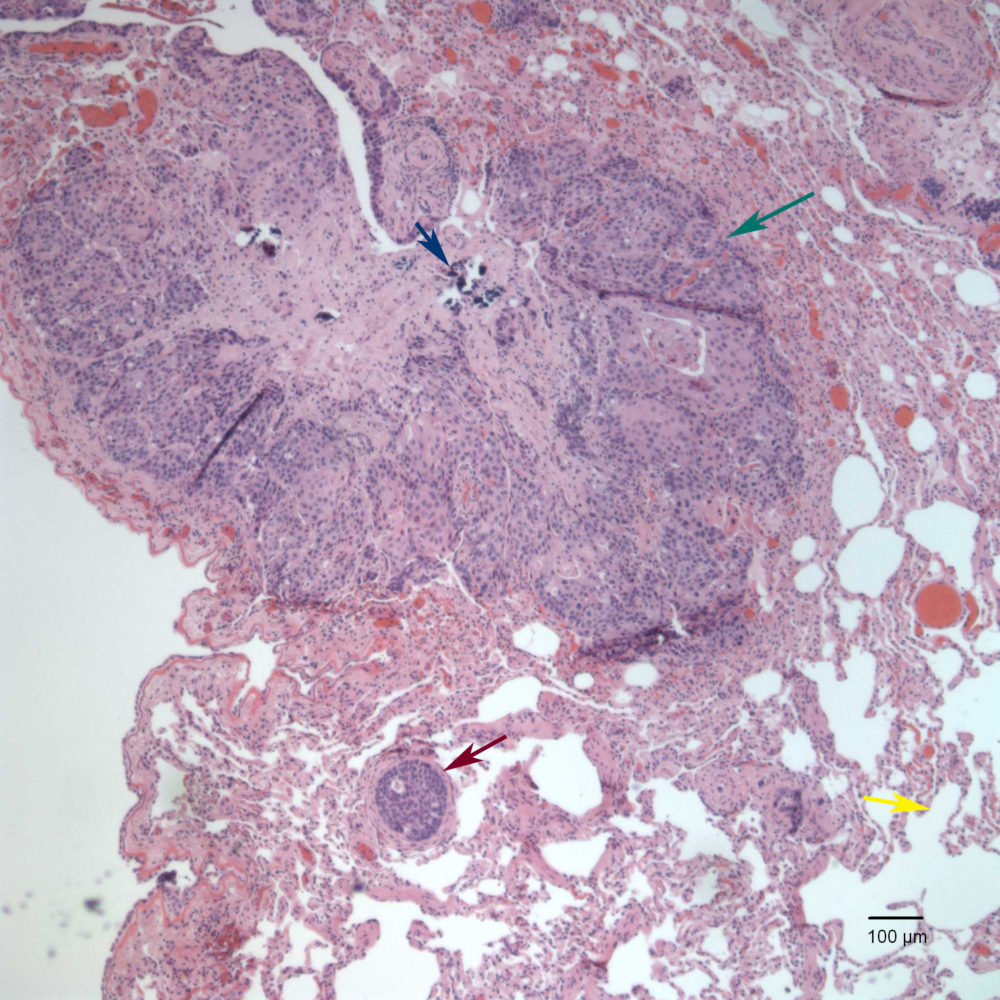
Expanding the lung parenchyma there is a well-defined, non-encapsulated neoplasm (green arrow) comprised of epithelial cells. Vascular invasion (red arrow) was present. The yellow arrow shows normal lung architecture. The blue arrow shows mineralized material compatible with dystrophic calcification (H&E, 4x objective)