
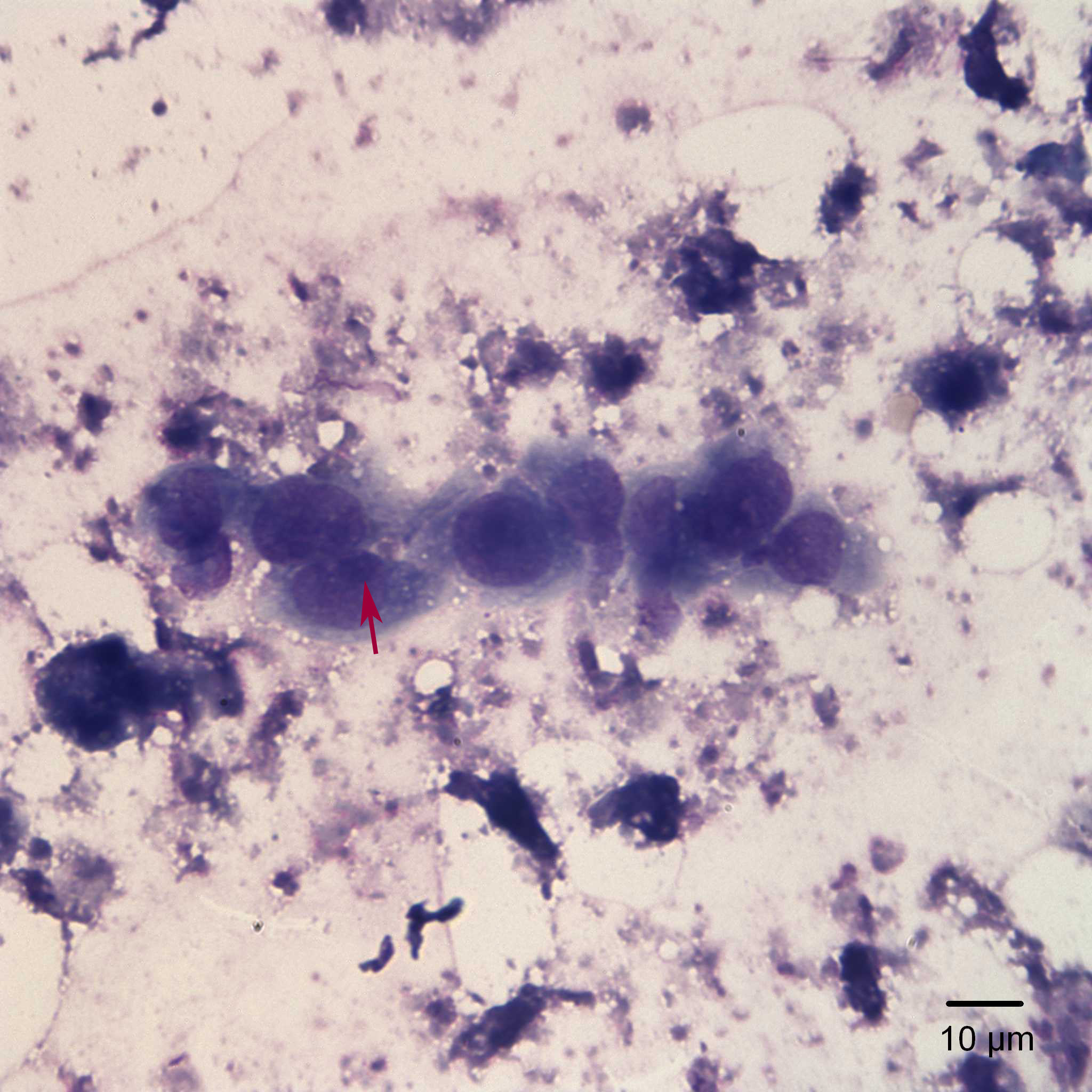
Fine needle aspirate of renal mass (50x objective, Wright’s stain): Smaller clusters of epithelial cells were also present. In this image, the nucleoli (red arrow) were more prominent and the nuclear chromatin was finer when compared to the bigger clusters shown in the previous image.