
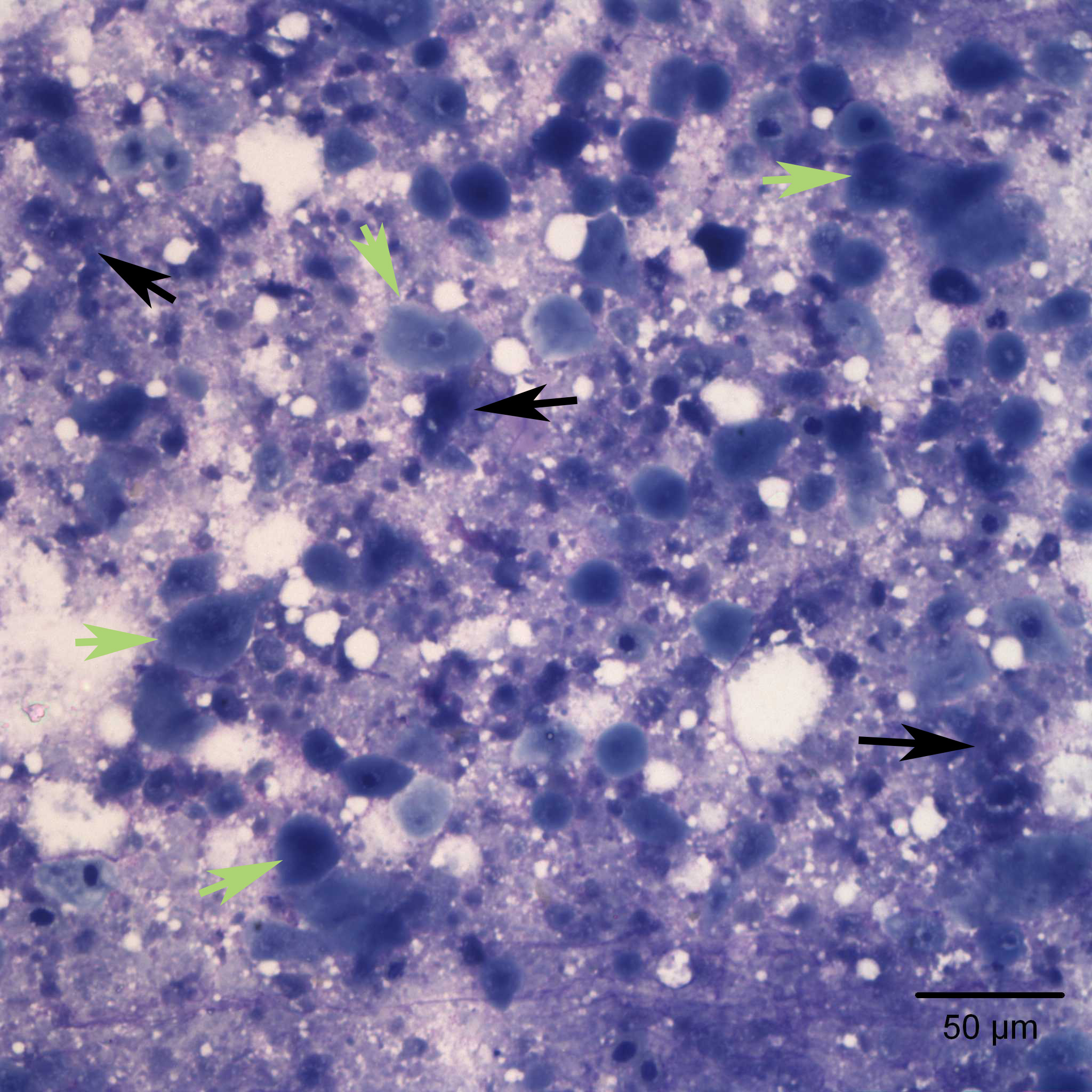
Innumerable fully keratinized squamous epithelial cells (green arrows) displaying nuclear to cytoplasm asynchrony on a background of necrosis (black arrows). The necrosis has a particular purple/gray muddy appearance on a Wright’s stain. Fading necrotic nuclei were often visible in the sample (20x objective)