
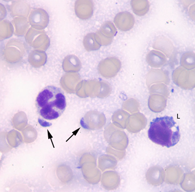
Figure 2b: Jejunal lymph node aspirate from a Wallaby. A single toxic neutrophil and reactive lymphocyte (L) are present in the bloody background, along with two zoites (arrows) (Wright’s stain, 1000x).
Figure 2b: Jejunal lymph node aspirate from a Wallaby. A single toxic neutrophil and reactive lymphocyte (L) are present in the bloody background, along with two zoites (arrows) (Wright’s stain, 1000x).
eClinpath helped 1.2 million visitors last year from 220 countries find important information on animal health. If you enjoy the site, please support our mission and consider a small gift to help us keep pace with its rapid growth. You can donate securely via PayPal or credit card. Thank you!
![]()