
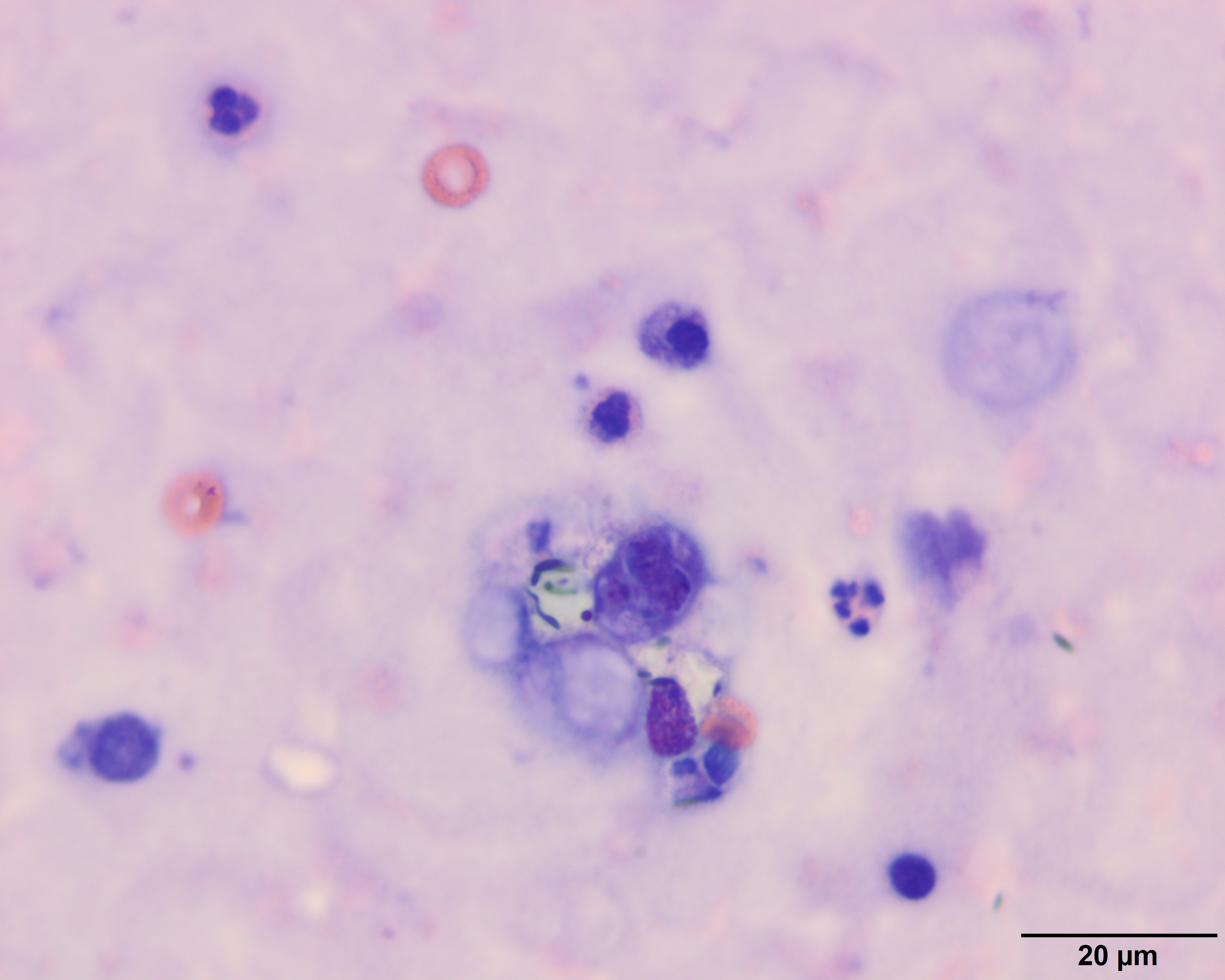
A sporulating dark blue Prototheca organism (just below center) with two adjacent empty casings and inflammatory cells (neutrophils, macrophages). A few erythrocytes and another empty casing (upper right) are in the background (Wright’s stain, 100x objective).