
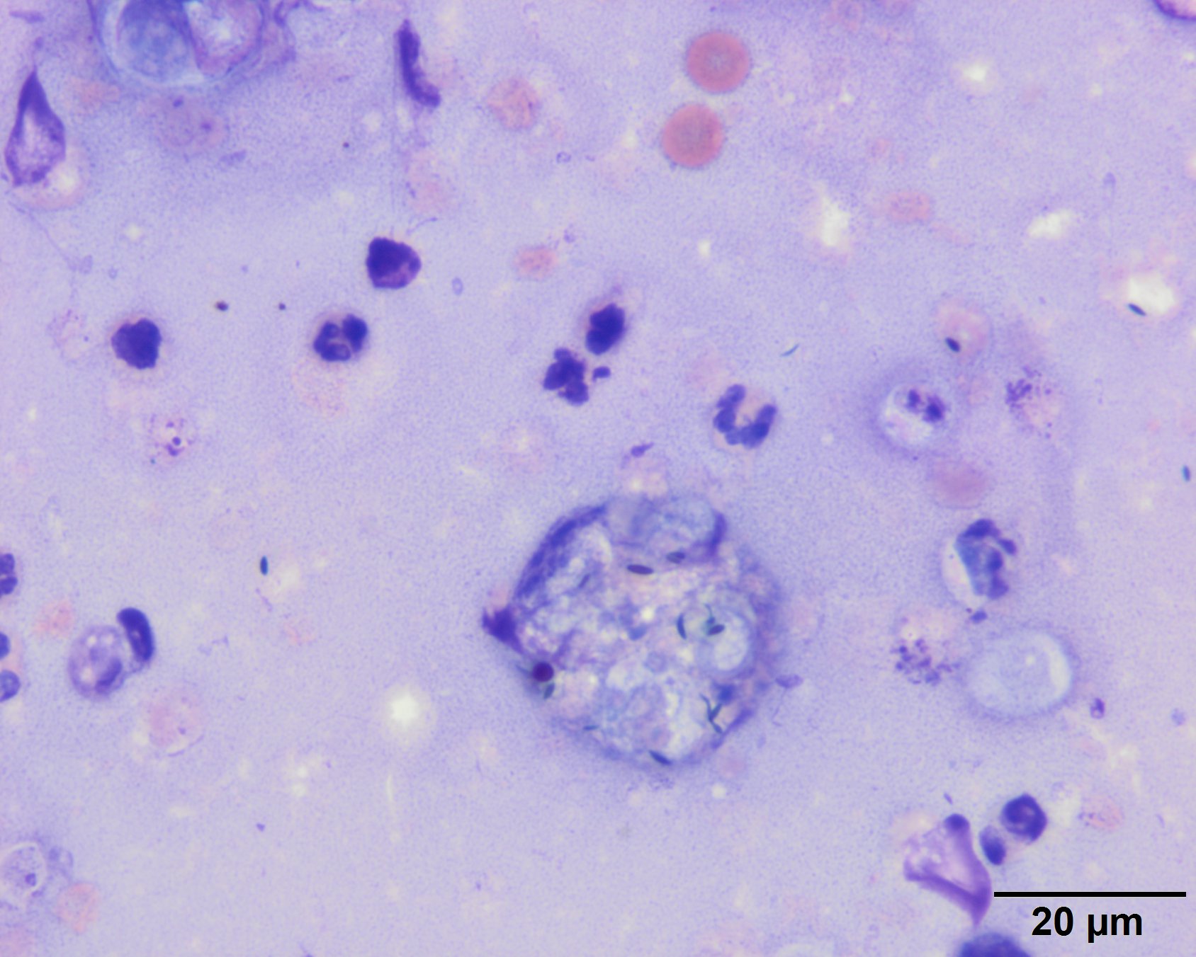
A macrophage (central) with phagocytized needle-like melanin granules, from the pigmented retinal epithelium, is present, along with an empty light blue casing (to the right of the macrophage). Neutrophils and erythrocytes are seen scattered in the background (Wright’s stain, 100x objective).