
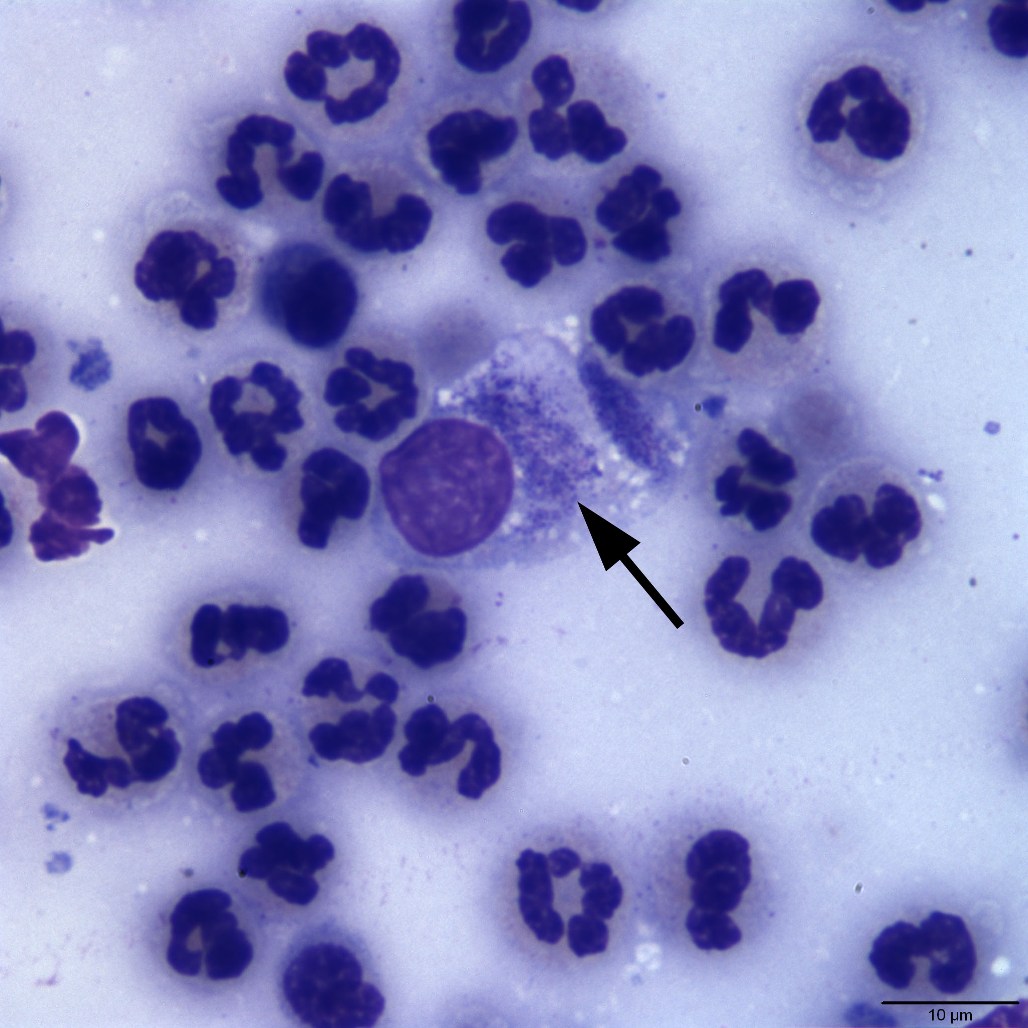
Representative high power image of a conjunctival swab from a cat. There is an epithelial cell in the center of the image. The arrow is pointing towards the intra-epithelial inclusions, which are suggestive of numerous elementary bodies of Chlamydia. If inclusions of this appearance were all that were seen, a valid differential diagnosis would be Mycoplasma felis organisms. Non-degenerate neutrophils and rare small lymphocytes are also in the field surrounding the epithelial cell. (Wright’s stain, 100x)