
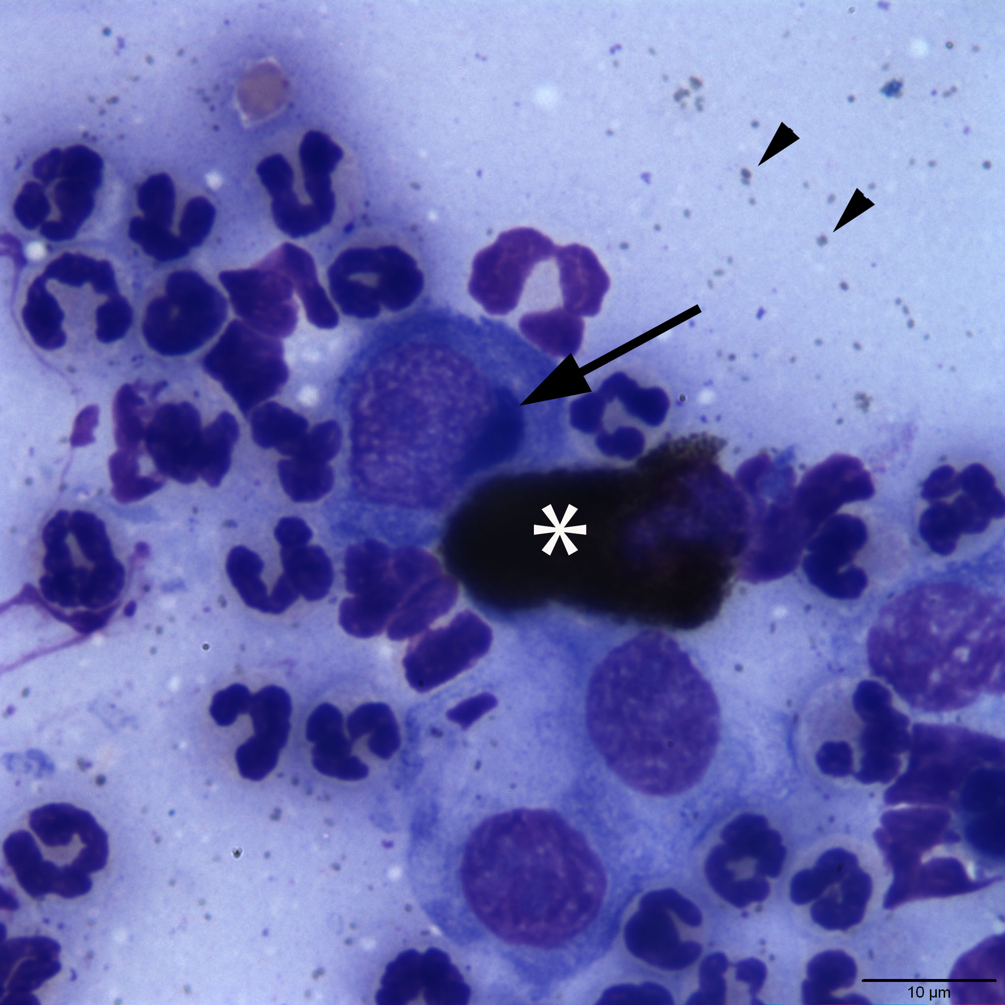
Representative high power image of a conjunctival swab from a cat. The arrow is pointing to a perinuclear inclusion consistent with Chlamydia within a conjunctival epithelial cell. The inclusion likely represents a reticulate body (or initial body, see Discussion). The adjacent cell (asterisk) is a heavily pigmented epithelial cell. Free melanin granules are also present throughout the background (arrowheads). Non-degenerate neutrophils are also in the image admixed with the epithelial cells. (Wright’s stain, 100x)