
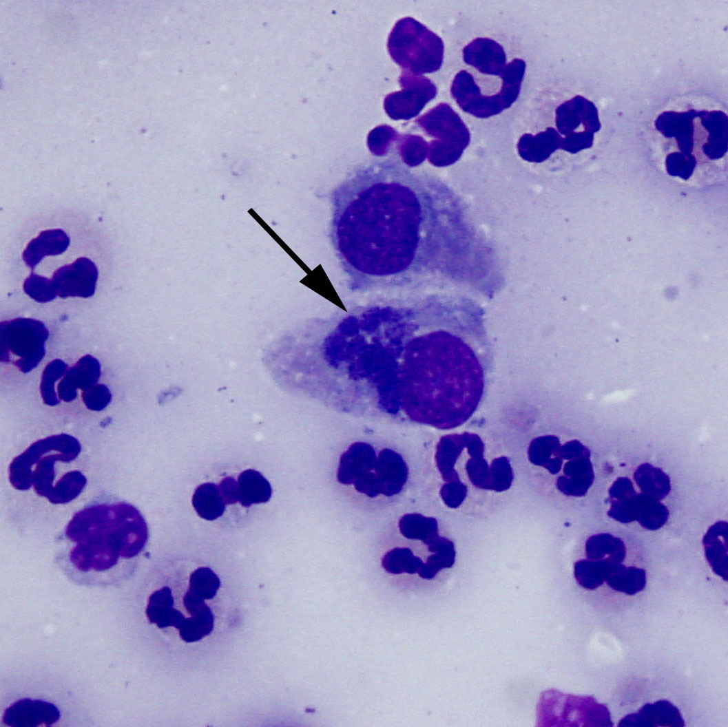
Representative high power image of a conjunctival swab from a cat. Two conjunctival epithelial cells are present in the center of the image. The arrow is pointing to the intracytoplasmic, perinuclear inclusions consistent with Chlamydia. These inclusions likely represent multiple reticulate bodies. The particulate appearance is likely due to the multiple elementary bodies within the reticulate body. Non-degenerate neutrophils are surrounding the epithelial cells. (Wright’s stain, 100x)