
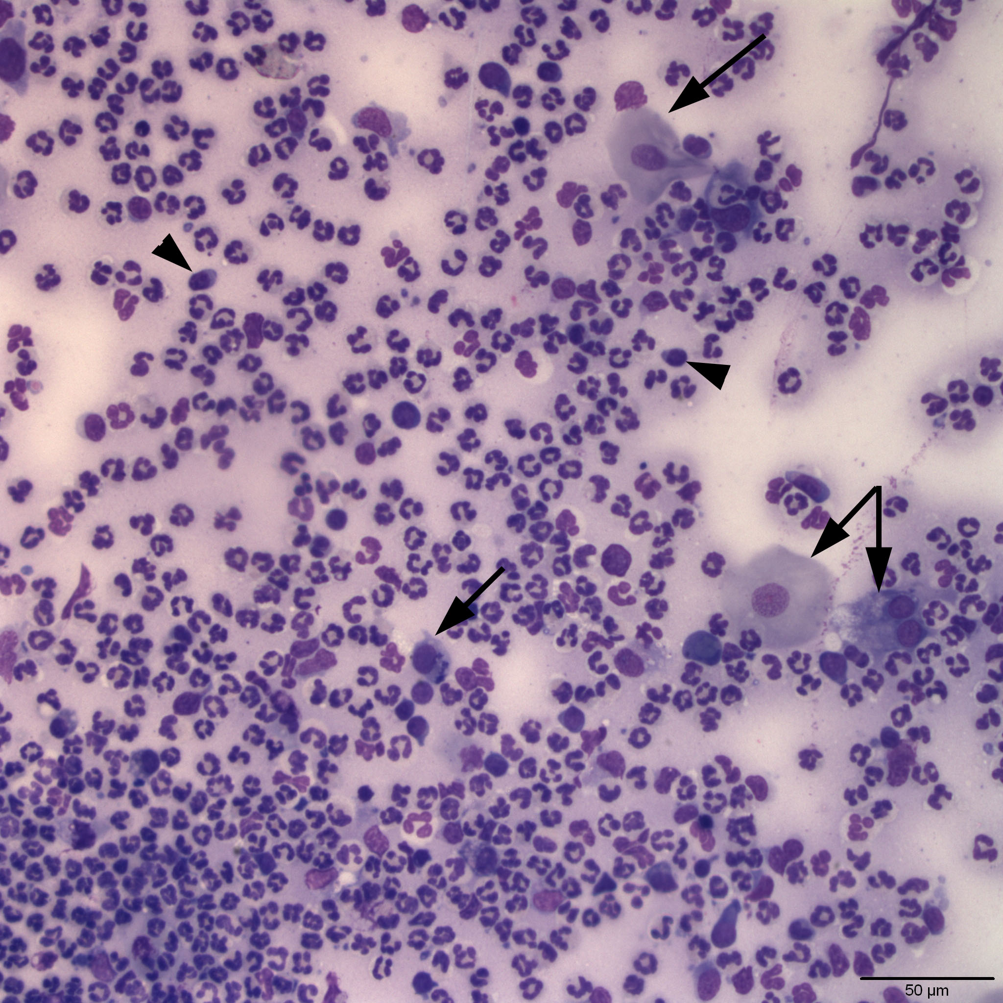
Representative low power image of a conjunctival swab from a cat. Note the numerous non-degenerate neutrophils throughout the field indicating marked neutrophilic inflammation. Low numbers of small lymphocytes are also present (arrowheads). Squamous epithelial cells are indicated by the arrows. A proteinaceous background is also evident by the amorphous purple hue most notable when looking around the peripheral margins of the cellular areas of the smear. (Wright’s stain 20x).