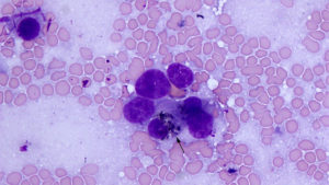
Scraping prepared from a biopsy of a liver mass in a horse: A cell with green-brown cytoplasmic pigment, similar to that seen in adjacent more mature hepatocytes (see Figures 3 and 3a), is evident in a presumptive tumor cell that is sandwiched between other tumor cells (arrow). This cytologic finding suggested the tumor cells were an immature variant of a hepatocyte. Note also the acinar-like arrangement (50x objective, Wright’s stain).

