
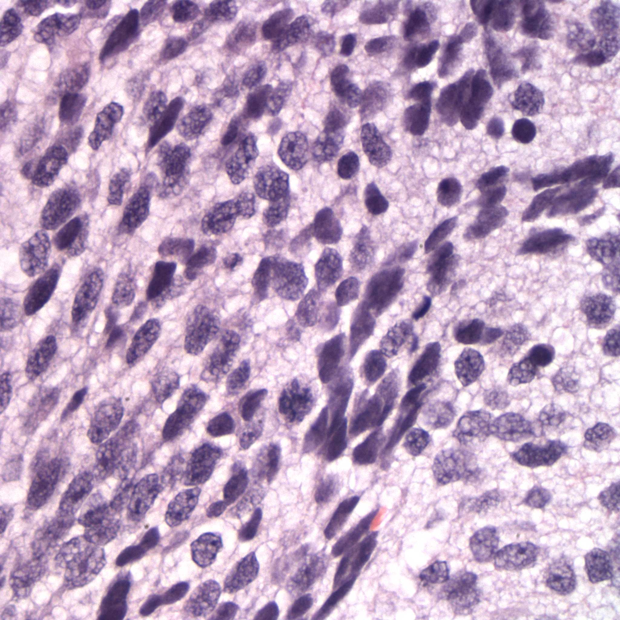
Figure 5. The normal cerebral parenchyma is markedly infiltrated and effaced by sheets of neoplastic cells. Neoplastic cells have indistinct margins and moderate amounts of vacuolated amphophilic cytoplasm. The nuclei are round to oval or irregularly shaped with densely clumped chromatin. Anisocytosis and anisokaryosis are moderate to marked (H&E stain).