
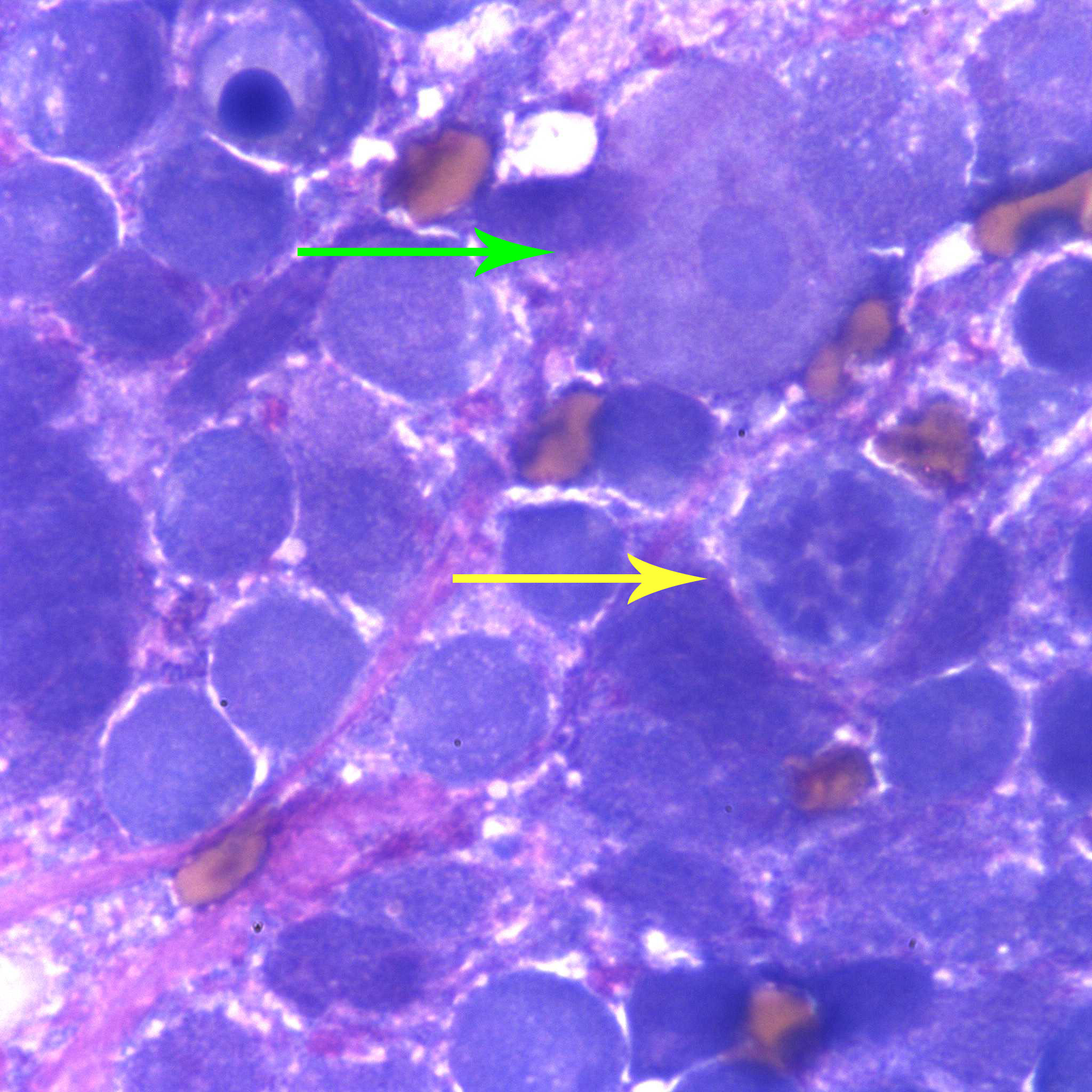
Figure 3a. The neoplastic cells are mostly individualized and have round to indented to pleomorphic (deeply lobulated) nuclei with finely stippled chromatin and small amounts of deep blue granular cytoplasm. The cells exhibit moderate to marked anisokaryosis and anisocytosis (green arrow illustrates a cell with a smaller nucleus and more abundant cytoplasm than adjacent cells). Mitotic figures are also seen (yellow arrow) (Wright’s stain, 1000x).