
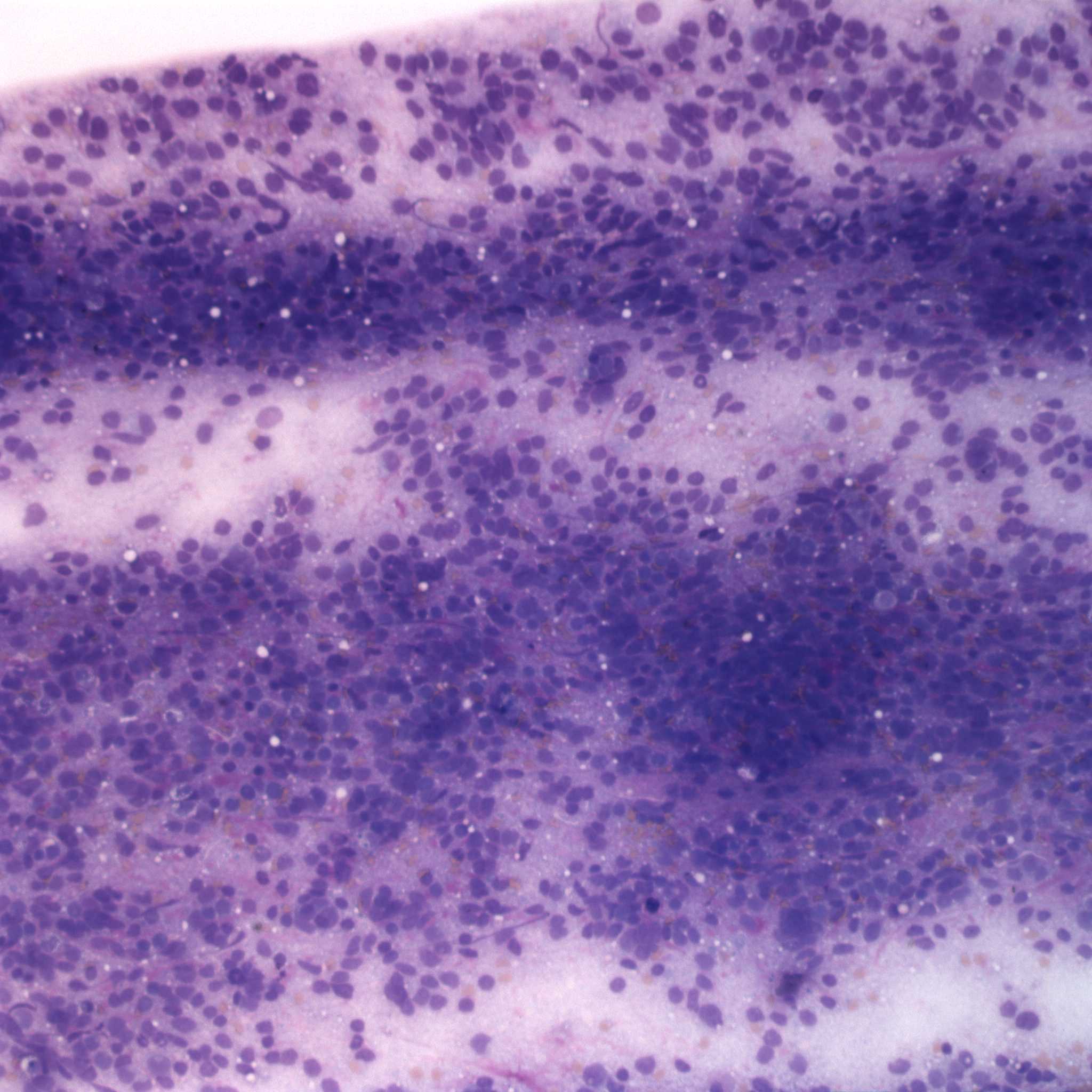
Figure 1. The smear is markedly cellular, consisting of individualized and densely packed cells with many ruptured cells seen as bare nuclei (Wight’s stain, 100x).
Figure 1. The smear is markedly cellular, consisting of individualized and densely packed cells with many ruptured cells seen as bare nuclei (Wight’s stain, 100x).
eClinpath helped 1.2 million visitors last year from 220 countries find important information on animal health. If you enjoy the site, please support our mission and consider a small gift to help us keep pace with its rapid growth. You can donate securely via PayPal or credit card. Thank you!
![]()