
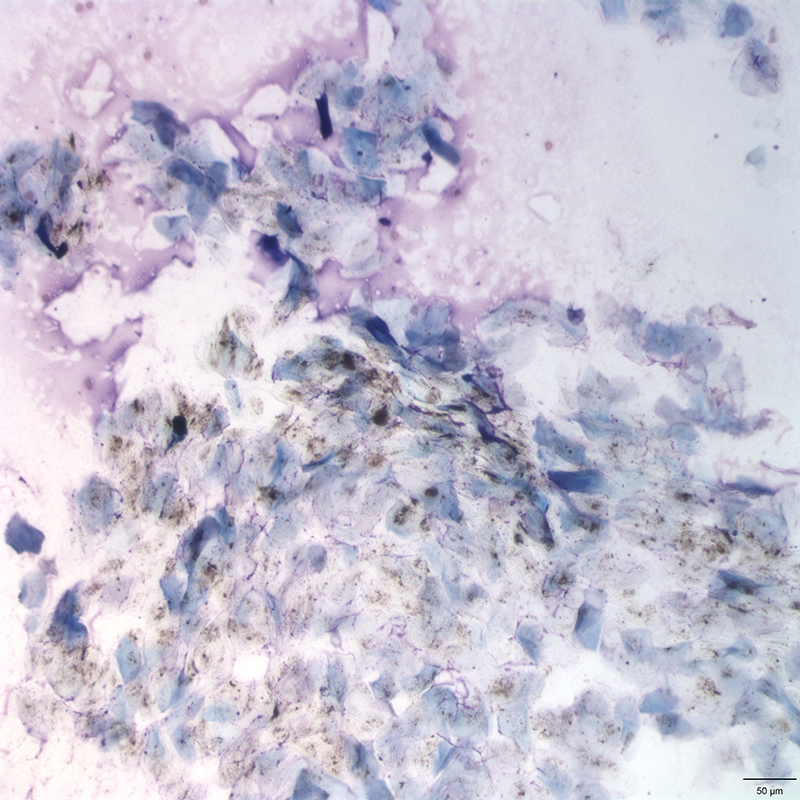
This image displays the typical cytologic findings from an aspirate of an intact follicular cyst. Note the abundant amount of anucleated keratinized squamous epithelial cells and keratin debris (some of which contains melanin granules) without evidence of inflammation.