
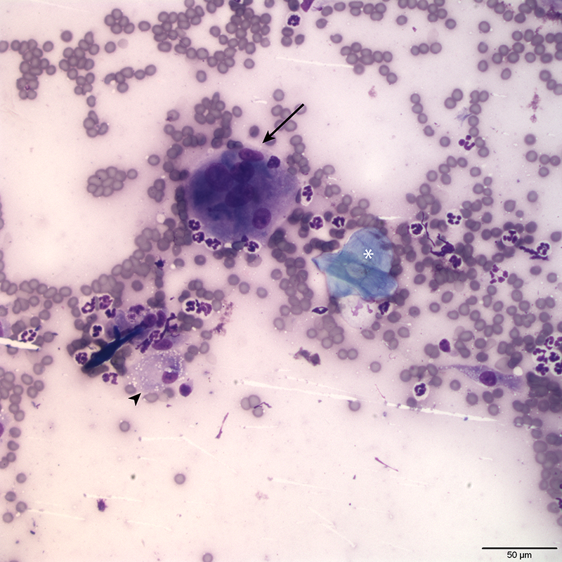
The inflammatory cells within the aspirate include neutrophils, macrophages (arrowhead), and occasional multinucleated macrophages (arrow). A keratinized squamous epithelial cell (asterisk) is also present in this field. The cell appears to have a central “clear” area corresponding to a faded nucleus and may be a so-called ghost cells. This suggests that the underlying cyst could be a matrical cyst, which is more likely to have ghost cells.