
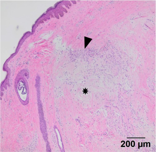
This image shows a portion of the neoplastic infiltrate. Note the neoplastic spindle cell population (arrowhead) expanding the dermis and elevating the overlying epidermis. The mass itself is unencapsulated, poorly cellular, and composed of spindle cells in interlacing bundles supported by abundant amounts of pale staining mucinous matrix material (*). Note the strands of collagen fibrils within the mass; these are particularly evident on the lower aspects of the mass (H&E stain, 4x objective).