
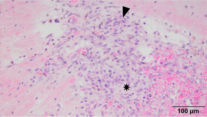
This higher power view shows the spindle cells (arrowhead) forming interlacing streams, embedded within light blue mucinous matrix (*). Note the area of hemorrhage with fibroblasts to the right of the asterix (H&E stain, 20x objective).
This higher power view shows the spindle cells (arrowhead) forming interlacing streams, embedded within light blue mucinous matrix (*). Note the area of hemorrhage with fibroblasts to the right of the asterix (H&E stain, 20x objective).
eClinpath helped 1.2 million visitors last year from 220 countries find important information on animal health. If you enjoy the site, please support our mission and consider a small gift to help us keep pace with its rapid growth. You can donate securely via PayPal or credit card. Thank you!
![]()