
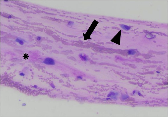
Note the windrowing (lining up) of red blood cells (arrow), individualized spindle cells within the matrix (arrowhead), and long thin fibrillar strands of pink to light purple mucinous matrix material (*) (Wright’s stain, 50x objective)
Note the windrowing (lining up) of red blood cells (arrow), individualized spindle cells within the matrix (arrowhead), and long thin fibrillar strands of pink to light purple mucinous matrix material (*) (Wright’s stain, 50x objective)
eClinpath helped 1.2 million visitors last year from 220 countries find important information on animal health. If you enjoy the site, please support our mission and consider a small gift to help us keep pace with its rapid growth. You can donate securely via PayPal or credit card. Thank you!
![]()