
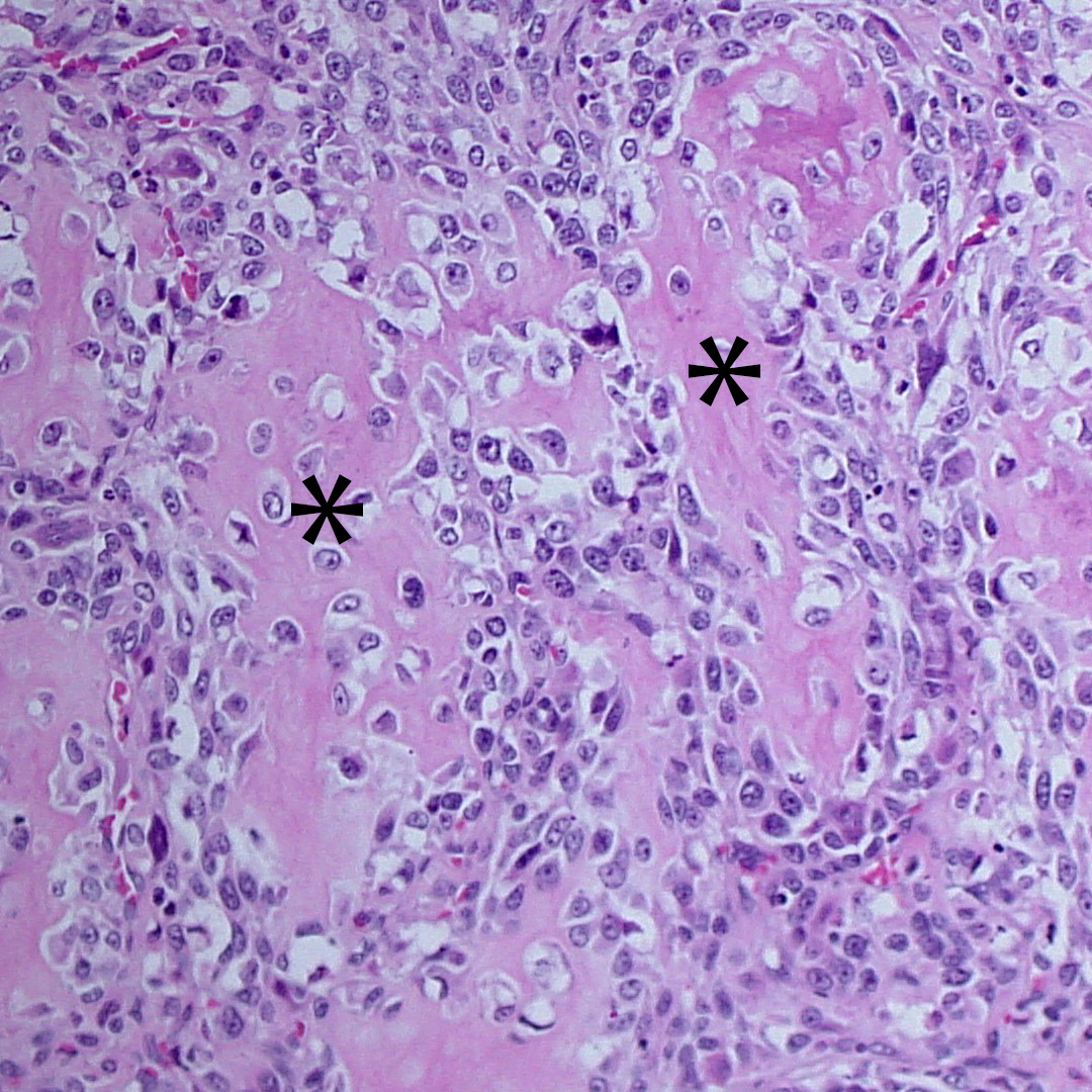
Figure 5. The tumor is comprised of many spindle cells (showing features of malignancy) that are surrounding islands of bright pink osteoid matrix (asterix) (H&E stain, 400x magnification).
Figure 5. The tumor is comprised of many spindle cells (showing features of malignancy) that are surrounding islands of bright pink osteoid matrix (asterix) (H&E stain, 400x magnification).
eClinpath helped 1.2 million visitors last year from 220 countries find important information on animal health. If you enjoy the site, please support our mission and consider a small gift to help us keep pace with its rapid growth. You can donate securely via PayPal or credit card. Thank you!
![]()