
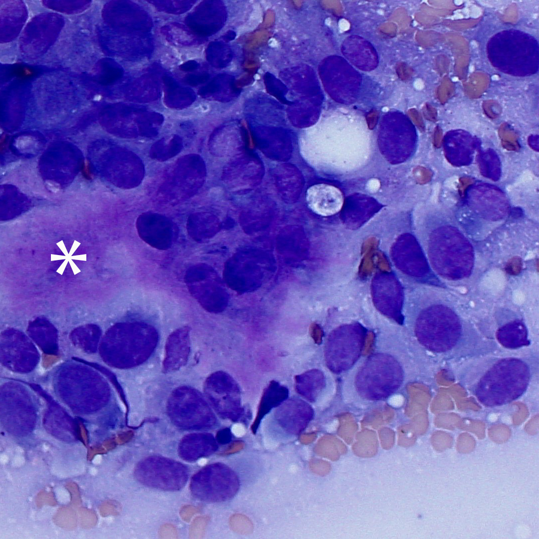
Figure 3a. Representative high power image of smears from a bone aspirate in a dog (Wright’ stain, 500x magnification). The smear contains many neoplastic spindle cells that are displaying numerous cytologic criteria of malignancy (such as moderate to marked anisokaryosis). The cells are surrounding a hot pink chunky to fibrillar extracellular matrix, most compatible with osteoid (asterix).