
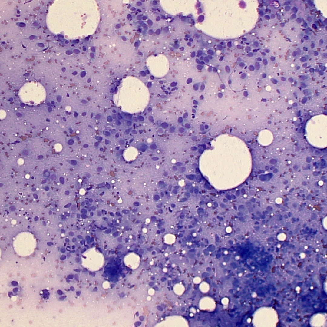
Figure 1a. Representative image from smears of a bone aspirate from a dog (Wright’s stain, 100x magnification). The smear is of high cellularity and consists of many individual to aggregates of spindle cells with free lipid and low numbers of erythrocytes.