
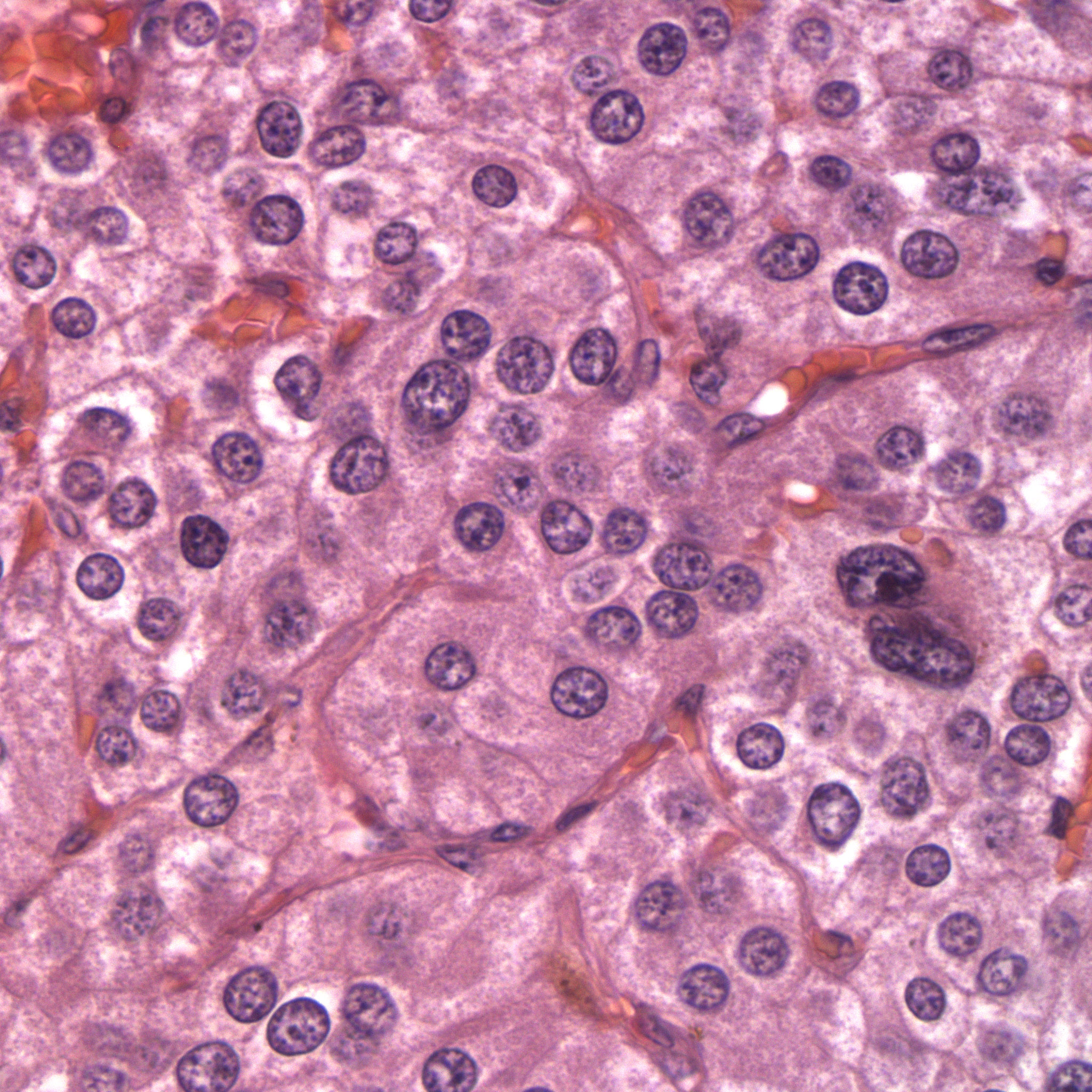
Figure 4: The neoplasm is composed of packets and cords of neoplastic polygonal cell surrounded by a fine fibrovascular connective tissue. The cytoplasm has a grainy texture. Some cells with very large nuclei are seen. (HE stain, 500x)
Figure 4: The neoplasm is composed of packets and cords of neoplastic polygonal cell surrounded by a fine fibrovascular connective tissue. The cytoplasm has a grainy texture. Some cells with very large nuclei are seen. (HE stain, 500x)
eClinpath helped 1.2 million visitors last year from 220 countries find important information on animal health. If you enjoy the site, please support our mission and consider a small gift to help us keep pace with its rapid growth. You can donate securely via PayPal or credit card. Thank you!
![]()