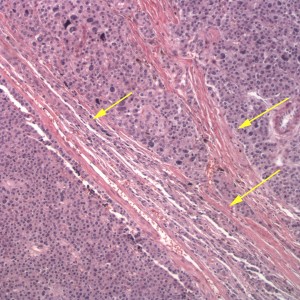
Figure 3: Neoplastic cells are seen in packets or nests separated by small amounts of fibrovascular stroma. The neoplastic cells infiltrate through the capsule (arrows), indicating malignant behavior, which is a feature of these tumors. (HE stain, 100x)

