
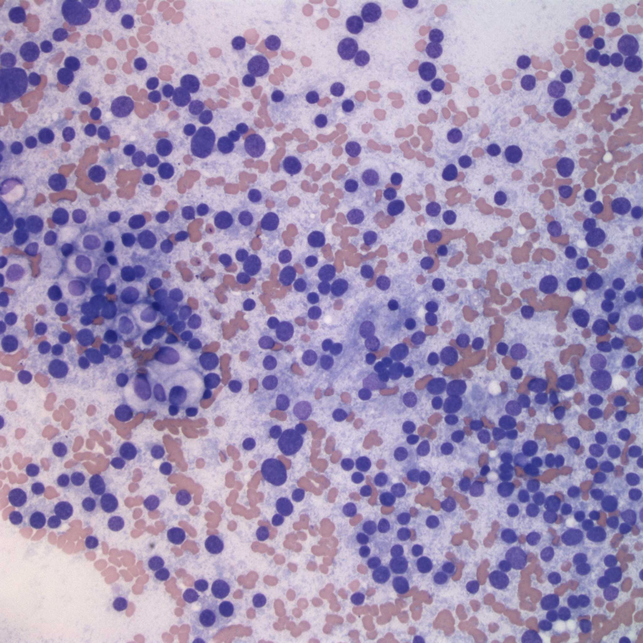
Figure 1: A monomorphic population of bare nuclei are dispersed in a pale, slightly granular purple background due to ruptured cytoplasm. Rare intact cells with pale cytoplasm are seen in amongst the rupturee cells. (Wright’s stain, 200x)
Figure 1: A monomorphic population of bare nuclei are dispersed in a pale, slightly granular purple background due to ruptured cytoplasm. Rare intact cells with pale cytoplasm are seen in amongst the rupturee cells. (Wright’s stain, 200x)
eClinpath helped 1.2 million visitors last year from 220 countries find important information on animal health. If you enjoy the site, please support our mission and consider a small gift to help us keep pace with its rapid growth. You can donate securely via PayPal or credit card. Thank you!
![]()