
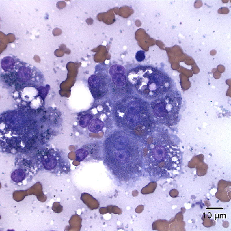
Two small clusters of hepatocytes. Some of the hepatocytes are binucleate and contain clear discrete cytoplasmic vacuoles compatible with lipid. There is also a small amount of blue-green cytoplasmic pigment, which could be lipofuscin or bile (no bile casts in this image). In the lower right corner, a 4-5 um long by 1 um wide protozoal zoite is present (Wright’s stain).