
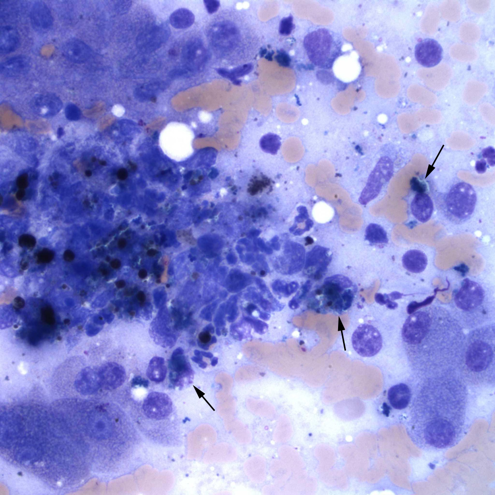
There is a dense aggregate or streak of partially ruptured difficult to discern cells between clusters of hepatocytes, with interspersed globules of bile pigment and increased numbers of inflammatory cells. The inflammatory cells are a mixture of non-degenerate neutrophils and macrophages (arrows), with fewer lymphocytes. The macrophages contain cytoplasmic pigment, which is likely a combination of hemosiderin, lipofuscin and copper (although the latter is not readily discernible) (Wright’s stain, 50x objective). We have associated this streaky cytologic pattern in liver aspirates with fibrosis (mast cells are often associated with these aggregates, but are not evident here). Note, the globules of bile pigment do not confirm cholestasis; linear casts need to be seen for that cytologic interpretation.