
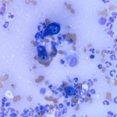
Figure 3: Several Blastomyces dermatitidis yeast (asterisks) illustrating the variable morphology of the organism. The yeast are distributed amongst a background of inflammatory cells consisting of neutrophils with moderate macrophages (Wright’s stain, x50 objective).