
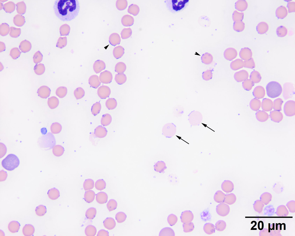
This higher power view illustrates many Mycoplasma organisms in ring and rod shapes on the red blood, usually on the edge but also on the surface (black arrowheads). There are several ghost cells (2 examples are illustrated with the black arrows) that have a lighter pink color and have organisms still attached. There are 2 polychromatophilic cells. Spherocytes are smaller cells (upper right corner) than more mature cells but are difficult to conclusively identify in cats (Wright’s stain).