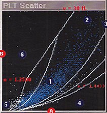
High light scatter (refractive index or internal complexity) is plotted as the X axis and low light scatter (cell size) is plotted as the Y axis (B). Platelets are detected in the region labeled 1. Large platelets (section 2) are identified on the basis of size (> 20 FL) and refractive index (which distinguishes them from red cells).

