
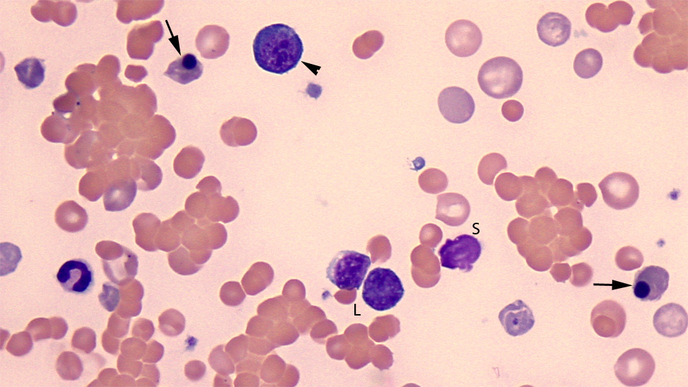
This image depicts two metarubricytes, with a dying pyknotic nucleus (that is about to be extruded) and polychromatophilic (purple) cytoplasm (arrows). There is also an earlier nRBC, which is a basophilic rubricyte (arrowhead), that has a deep blue cytoplasm and round nucleus with clumped chromatin. Contrast both types of nRBC to the two lymphocytes (L) in the smear, which have a lighter chromatin and less blue cytoplasm (although the cell on the right is difficult to tell from a nRBC). There is also a smudged cell (S, identity uncertain) (Wright’s stian, 100x objective). Note the agglutination and spherocytes (small cells that lack central pallor) and evidence of regeneration (polychromasia).