
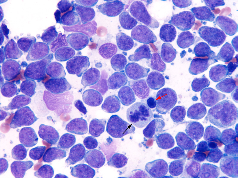
Large lymphocytes with fine chromatin, multiple 1-3 peripheral nucleoli and small amounts of deep blue grainy cytoplasm dominate in this lymph node aspirate (Wright’s stain, 500x magnification). A single mitotic figure is noted (black arrow). The large size of the cells can be discerned in relation to a small mature lymphocyte (with clumped or smooth chromatin) (red arrow). The tumor cells were positive for Pax-5 on immunohistochemical staining, indicating a B cell lymphoma. Low numbers of lymphoblasts were observed in a peripheral blood smear (with no cytopenias) and the dog had <5% lymphoblasts in a bone marrow aspirate.