
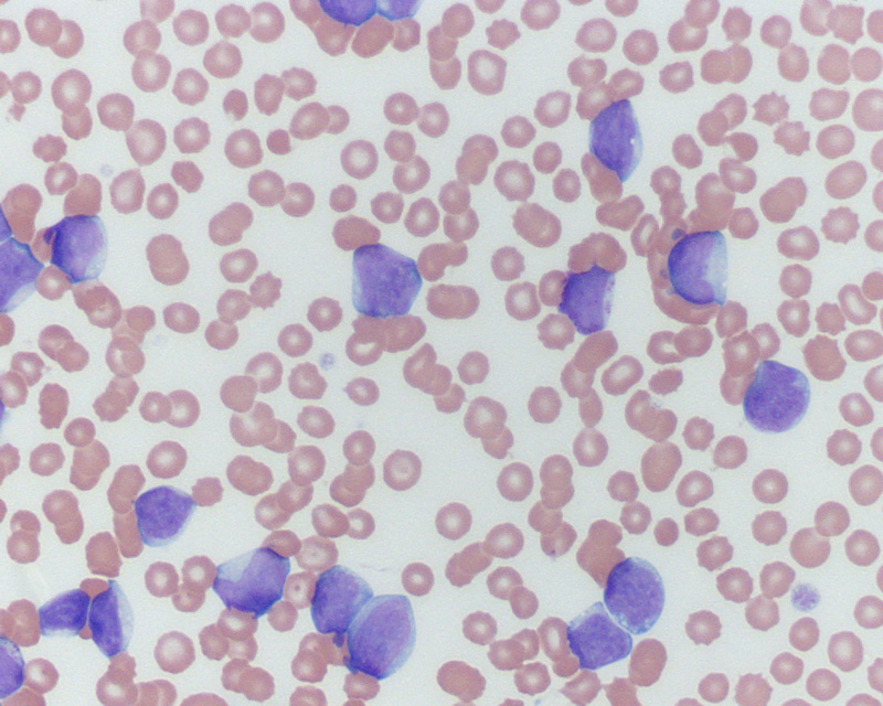
This dog had a marked leukocytosis (300,000/uL) consisting of large blasts with a concurrent pancytopenia (non-regenerative anemia, neutropenia, severe thrombocytopenia. Some of the cells had coarse red-purple granules. A few eccentrocytes are evident, indicating a degree of oxidant injury (not uncommon in dogs with cancer) (Wright’s stain, 500x magnification). This indicates an acute leukemia, which could be myeloid or lymphoid. A bone marrow aspirate confirmed this diagnosis (see related image). To distinguish between these two main categories of leukemia, immunophenotyping was performed. The tumor cells were positive for the stem cell markers, CD34 and CD90, and for markers expressed on monocytes and some T lymphocytes (CD1c) but were negative for MHCII. Unfortunately, they lacked other differentiation markers (including other T cell markers, such as CD3 and CD5), precluding a definitive diagnosis by immunophenotyping. Cytochemical staining was then performed and the tumor cells were strongly positive for leukocyte alkaline phosphatase, a marker of monoblasts in the dog. This supported a final diagnosis of acute monoblastic leukemia, the most common type of AML in the dog in our experience.