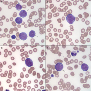
Venous blood smear from a dog with acute myelomonocytic leukemia (Wright’s stain, 100x objective). The diagnosis was based on immunophenotyping by flow cytometry (positive for CD34, CD4, CD11b and CD90 and negative for MHCII) and cytochemical staining (positive for ALP and ANBE): A: Myeloid blasts with cells differentiating towards monocytes (promonocytes due to their high nuclear to cytoplasmic ratio and fine chromatin, arrows). The small cell is a lymphocyte (arrowhead). B: A monoblast with a slightly irregular nuclear outline but fine chromatin and 2 prominent nucleoli. The nucleus is not lobulated enough to call a promonocyte, but in this variant of AML and acute monoblastic/monocytic leukemia, promonocytes are included in the blast count. C: This cell illustrates the difficulty in distinguishing blasts from promonocytes. The cell has fine chromatin and 4 distinct nucleoli but the nucleus is lobulated. Is it a blast or promonocyte? This determination probably does not matter in this type of leukemia because this cell would be included in the blast count. D: On first appearance the cell on the left (arrow) looks like a blast, but it has a lobulated nucleus – is it a promonocyte? This distinction is likely moot (see C above). The cell on the right is a blast and the remaining cell is a band neutrophil (arrowhead).

