
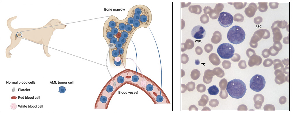
Left panel: In acute myeloid leukemia (AML), one or more genetic mutations in a hematopoietic stem cell causes proliferation of the neoplastic clone, outcompeting the residual normal hematopoietic cells (megakaryocytes, erythroid and myeloid progenitors). In many animals, the neoplastic cells spill into blood, resulting in a marked leukocytosis, comprised primarily of tumor cells with single or multiple cytopenias (non-regenerative anemia, neutropenia, thrombocytopenia) from decreased production of normal hematopoietic cells (neutrophils, red blood cells, platelets). (Created with Biorender.com.)
Right panel: In this representative image of a blood smear from a dog with AML and a marked leukocytosis, tumor cells dominate (white *), with low numbers of normal white blood cells (WBC; band neutrophil), red blood cells (RBC), and platelets (arrowhead). The dog had pancytopenia (non-regenerative anemia, neutropenia with a left shift and mild toxic change, and severe thrombocytopenia). Immunophenotyping and cytochemical staining confirmed an acute myeloid leukemia of monocytic origin (modified Wright’s stain, 100x objective).