
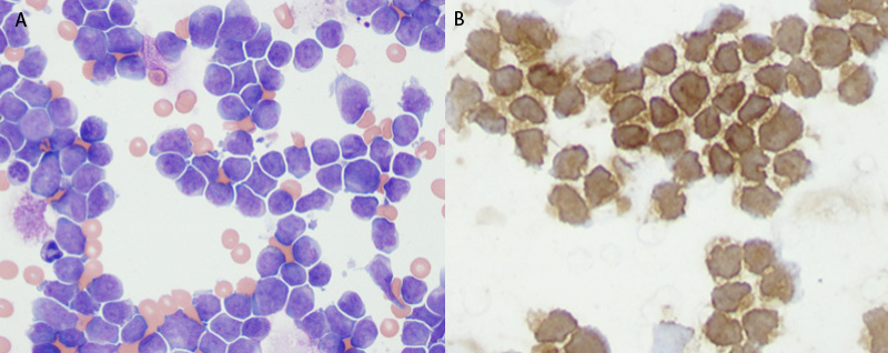
Immunocytochemical staining for T (CD3) and B (Pax-5) was done on cytospin smears prepared from a lymph node aspirate from a dog with multicentric lymphoma. A: Intermediate lymphocytes with clefted to deeply cleaved nuclei, no nucleoli, fine chromatin and scant amounts of cytoplasm dominate (Wright’s stain). B: The cells showed strong membraneous reactivity for CD3, indicating a T cell lymphoma (CD3 immunostain). No circulating cells were seen in this dog and none of the tumor cells were positively identified in a marrow aspirate. This was classified as an indolent T zone lymphoma (low grade intermediate cell) on histologic examination.