
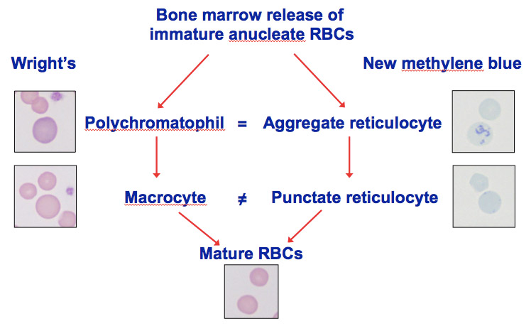
There are two types of immature a nucleate RBC, which have a different appearance on Romanowsky (polychrome)-stained smears (Wrigth’s stain or quick stains, such as Diff-Quik) and new methylene blue-stained smears (new methylene blue binds to and precipitates the RNA in these immature RBC).
1) Aggregate reticulocytes in a new methylene blue-stained smear correspond (=) to polychromatophils in a Wright’s-stained smear (i.e. all polychromatophils are reticulocytes). Aggregate reticulocytes are more immature (contain more RNA) than punctate reticulocytes and mature into the latter, in the circulation or in the bone marrow. In the cat, this takes 12 to 24 hours.
2) Punctate reticulocytes in a new methylene blue-stained smear can be (but are not always, ≠) macrocytes (larger red cells) in a Wright’s-stained smear. They can also be of normal size. Whether they are macrocytic or normocytic, punctate reticulocytes stain red in a Wright’s-stained smear (they have too little RNA to stain purple). Punctate reticulocytes are more mature (contain less RNA) than aggregate reticulocytes and are remodeled as they circulate in the bloodstream to mature RBC. Note, just as not all punctate reticulocytes are macrocytes, not all macrocytes are punctate reticulocytes. Macrocytes can be formed through other mechanisms, including abnormal DNA metabolism (e.g. myelodysplastic syndrome). Such macrocytes may lack RNA and will not be identified as punctate reticulocytes in a new methylene blue-stained smear. Equidae release macrocytes (a few of which are indeed punctate reticulocytes) and very rarely do not release aggregate reticulocytes (polychromatophils) in a regenerative anemia. Similarly cats with mild anemia may only release punctate reticulocytes.