
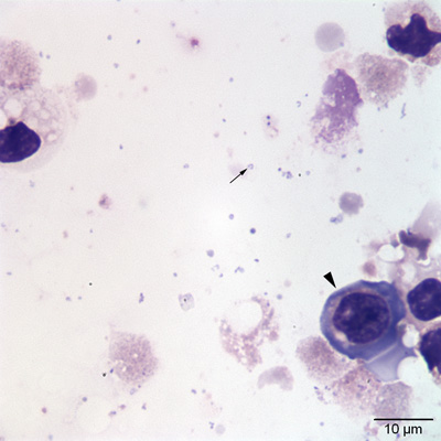
Figure 3: Individual small ring-shaped (arrow) to coccoid bacteria are seen in the background. The epithelial cell in this image (arrowhead) has a higher nuclear to cytoplasmic ratio than the others represented in Figures 1 and 2. This variation is attributed to inflammation-induced hyperplasia or dysplasia (Wright’s stain).