
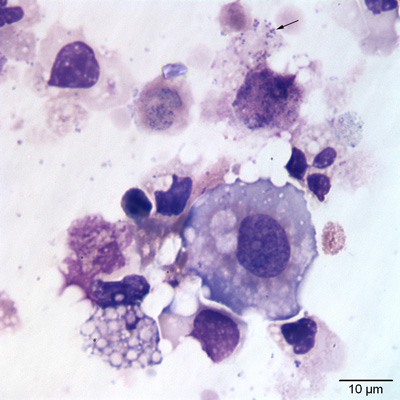
Figure 2: A large epithelial cell alongside a vacuolated macrophage, dying cells and swollen neutrophils is evident. Numerous coccoid small bacteria are seen in the one ruptured cell and some of the bacteria are also present in the background (Wright’s stain).