
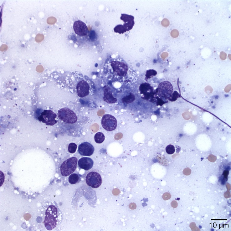
Here there are mixed inflammatory cells, mostly comprised of macrophages and fewer small lymphocytes. Some of the cells are smudged and cellular outlines are indistinct on some macrophages (Wright’s stain, 50x)
Here there are mixed inflammatory cells, mostly comprised of macrophages and fewer small lymphocytes. Some of the cells are smudged and cellular outlines are indistinct on some macrophages (Wright’s stain, 50x)
eClinpath helped 1.2 million visitors last year from 220 countries find important information on animal health. If you enjoy the site, please support our mission and consider a small gift to help us keep pace with its rapid growth. You can donate securely via PayPal or credit card. Thank you!
![]()